Comprised primarily of water and collagen, the cornea is the clear outer layer of the eye which plays an essential role in vision and eye health. The cornea focuses light into the eye so you can see clearly and also acts as a protective outer layer against debris and germs. But what happens when it gets damaged? If the cornea becomes damaged through injury or infection, the resulting scaring or discoloration can can lead to vision loss.
Research has found that the use of collagen can promote epithelial and stromal healing within pets. Quick healing of the corneal surface is ideal for the comfort of the patient as well as for the prevention of infection and scaring.
What is an ulcer or an abrasion?
Corneal ulcers and abrasions are painful open wounds within the cornea. The most common symptoms are squinting, excessive blinking, holding the eye fully closed, redness and swelling of the sclera (the white part of the eye), the front of the eye becoming hazy or cloudy, or discharge from the eye.
Damage to the first few layers of the epithelium is known as a corneal abrasion. Corneal ulcers are a deeper erosion or injury through the epithelium and into the stroma layer. This form of injury can lead to fluid collection in the stroma, giving the pet’s eye a cloudy appearance. If the injury continues deeper into the anterior surface of the cornea – the Descemet’s membrane – a descemetocele is created. This is an area of extreme corneal thinning which can result to irreversible damage such as perforation, the rupturing of the Descemet’s membrane and collapsing of the eye itself.

How do corneal ulcers and abrasions come about?
Corneal ulcers are one of the most common eye diseases seen in dogs. Damage to the cornea can occur at any moment of time in animals of all ages. Because the injury is to the surface of the eye, anything from debris or roughhousing with other animals can lead to a scratch on the cornea. Some breeds are at a higher risk of corneal ulcerations.
Brachycephalic breeds such as Pugs and Persian cats are more likely to suffer from corneal ulcers. This is due to numerous factors such as the anatomy of their small skulls that increases the exposure of their eyes to the exterior environment, poor corneal sensitivity and a lessened blink coverage over the cornea. Other breeds that commonly suffer from ulcers of abrasions such as Boxers have inherited epithelial dystrophy or weaking of the cornea. Animals with Keratoconjunctivitis sicca (KCS) or tear deficiency are also predisposed to corneal ulcers due to the decreased tear production and increased drying of the cornea.
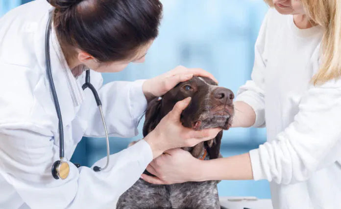
What can you do to promote corneal healing?
Treatment depends on the type of corneal injury. Corneal abrasions are the least serious, and generally heal within 3-5 days through medication such as ophthalmic antibiotic drops or ointment to prevent bacterial infections. If a corneal ulcer or a descemetocele is present, veterinarians must take additional steps to protect the eye and to promote healing. Following a surgical intervention such as a corneal graft, a collagen shield can be used to protect the ocular surface. Keep in mind that a collagen shield can be used for abrasions as well to ease the animal’s discomfort and promote healing.
Made of collagen proteins and shaped in the form of a contact lens, collagen corneal shields can be easily placed on the surface of the eye. They provide a protective barrier over the eye to support the regeneration of epithelial cells, which typically fill defects of the cornea within 48-72 hours. But how does the collagen corneal shield dissolve? The shield will start degrading by the presence of collagenases – naturally present in the animal’s tears – which are enzymes that degrade the collagen protein. Depending on how much collagen is in the shield, shields on the market will take between 12-72h to degrade. This method of healing corneal ulcers with collagen shields has been found to “enhance drug delivery, promote healing of corneal tissue and reduce corneal inflammation” (Willoughby et al, 2002).
What is the VET SHIELD™?
The cornea receives oxygen and nutrients from tears that are spread over the cornea when an animal blinks. The VET SHIELD™ is a bovine collagen-based shield that creates a biocompatible barrier designed to be placed over the cornea of a non-patched eye. Due to its biocompatibility, the shield lets oxygen pass through freely, while preventing the eyelid or other material from rubbing on the cornea. This collagen based corneal shield allows nutrients to reach the epithelial cell layer and encourages a more rapid recovery of corneal abrasions and ulcers with no pain to the patient. The VET SHIELD™ is also permeable to water-based medications (not ointments), thus not requiring the removal and replacement of the lens when medications are applied. This collagen based corneal shield begins to degrade after 72 hours and as the shield gradually dissolves, a thin layer of collagen is released which helps lubricate the eye.

The cornea not only protects the eye but a healthy cornea is essential for a pet to have normal vision. While corneal abrasions and ulcers can happen at any time, it’s key that veterinarians have innovative tools at their disposal to support the health and comfort of their pet patients. The VET SHIELD™ collagen corneal shield is an ideal solution for cats and dogs for their ulcers, abrasions and following eye surgery. The VET SHIELD™ encourages the natural healing of corneal injuries while protecting the eye from further damage.
References
Cleveland Clinic,(2018, February 14th). Cornea Transplant. My Cleveland Clinic. https://my.clevelandclinic.org/health/treatments/17714-cornea-transplant
Farghali, H.A., AbdElKader, N.A., AbuBakr, H.O., Ramadan, E.S., Khattab, M.S., Salem, N.Y. and Emam, I.A., 2021. Corneal Ulcer in Dogs and Cats: Novel Clinical Application of Regenerative Therapy Using Subconjunctival Injection of Autologous Platelet-Rich Plasma. Frontiers in Veterinary Science, 8, p.123.
Packer R. M.A, Hendricks A. Burn C.C., 2015. Impact of Facial Conformation on Canine Health: Corneal Ulceration. PLOS ONE, 10(5): e0123827. https://doi.org/10.1371/journal.pone.0123827
Simşek, N.A., Ay, G.M., Tugal-Tutkun, I., Başar, D. and Bilgin, L.K., 1996. An experimental study on the effect of collagen shields and therapeutic contact lenses on corneal wound healing. Cornea, 15(6), pp.612-616.
Startup, F.G., 1984. Corneal ulceration in the dog. Journal of Small Animal Practice, 25(12), pp.737-752.
Willoughby, C.E., Batterbury, M. and Kaye, S.B., 2002. Collagen corneal shields. Survey of ophthalmology, 47(2), pp.174-182.